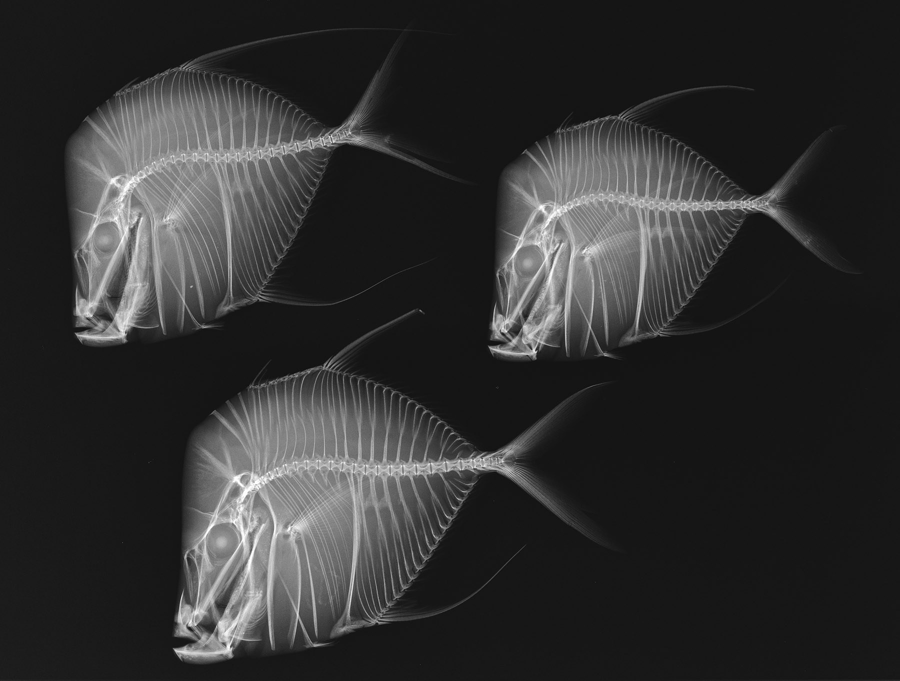

Selene vomer (Lookdown), one of the photographs in X-Ray Vision. Credit: Sandra J. Raredon, Division of Fishes, NMNH.
Superman has it, and so does the Smithsonian Institution: x-ray vision. We’ve just finished hanging 40 intriguing x-ray images of fish specimens from the USA’s National Fish Collection, a library of more than 4 million preserved specimens of some 20,000 fish species from around the world held by the Smithsonian Institution in Washington DC (that’s a lot of preserving jars).
These are a stark contrast to the full colour life size photos of whales in the Beautiful Whale exhibition that X-Ray Vision replaces in the USA Gallery. Being fish-sized they’re much easier to hang (the panels in Beautiful Whale were up to four metres long and two metres high each) and just as interesting.

Richard Wood during the installation of X-Ray Vision.
I’m not an ichthyologist and have a phobia about fish bones (I think it’s from being constantly warned as a child to watch out for bones in the freshly cooked catch we took from the Brisbane Water on school holidays), but the chance to look at fish inside out is another thing altogether.
Inside, fish appear incredibly fine and at times implausible, with individual bones ending in hair-width wisps of calcium. The species the Smithsonian’s curators Sandra Raredon and Lynne Parenti have selected to x-ray for the exhibition are intriguing; they range from the Pelican Eel to Dhiho’s seahorse.

Eurypharynx pelecanoides (Pelican Eel). Credit Sandra J. Raredon, Division of Fishes, NMNH.
The Pelican Eel trolls temperate and tropical oceans between 500 and 3,000 metres down using its huge mouth and pouched lower jaw to collect food. In our x-ray image you can see the last fish this specimen caught protruding from its stomach. The tiny, 25mm long seahorse only lives near Japan.
The exhibition is hung in a strict order from the least to most evolved fish species and each framed image has a label that includes a photo of the specimen that was x-rayed. Some, like the Bulbous Deep Sea Angler are extremely rare with only 3 specimens ever caught, one near Townsville.

Caroline from the conservation team installing works from A Different Vision.
X-rays have been used for medical and investigative imaging for just on 120 years, but Aboriginal and Torres Strait Islander artists have depicted the skeletons, muscles and organs of marine life for thousands of years.
We’re also installing A Different Vision, as a parallel exhibition in the USA Gallery. This is a bijoux collection of traditional and contemporary Aboriginal and Torres Strait Islander art using vision of different kind.
Explore the Smithsonian’s gallery of images on Flickr.